Mikroskopkamera: Zuverlässiger & präziser Autofocus
Veröffentlicht am 20. August 2016 von TIS Marketing.
Ursprünglich veröffentlichter Artikel in der Fachzeitschrift Mikroskopie (Jahrgang 3, Nr. 1/2016, S. 37-51) von J. Piper und M. Torzewski. Gegliedert in Abschnitt: 1, 2, 3, 4, 5, 6, 7, 8, 9, 10 und 11.
Autofokus
Der Autofokus der Kamera funktioniert überraschend zuverlässig und präzise. Auch in schwierigen Einstellungssituationen arbeitete dieser in allen praktischen Tests absolut zielsicher. Bei visueller Kontrolle ließ sich die vom Autofokus ermittelte Schärfeebene nicht weitergehend korrigieren bzw. verbessern. Auch wenn das Bild im Mikroskop selbst unscharf fokussiert wurde, konnte der Autofokus über weite Verstellbereiche das Monitorbild scharf fokussieren, ohne dass der Arbeitsabstand des Mikroskop-Objektivs verändert wurde. Eine exemplarische Messung des Verstellbereichs wurde mit einem Objektiv L 32/ 0.40 (mit langem Arbeitsabstand, kombiniert mit Okular 10x) durchgeführt. Wenn das beobachtete Präparat in Mittelstellung vom Autofokus scharf fokussiert war, konnte die Fokussierung am Mikroskop in beiden Richtungen um 8 µm verändert werden, ehe der Autofokus das im Mikroskop unscharf fokussierte Bild nicht mehr auf dem Bildschirm nachfokussieren konnte. Somit konnte der Autofokus bei diesem Objektiv insgesamt einen vertikalen Verstellbereich von 16 µm kompensieren. Dies entspricht etwa dem Dreifachen der visuell wahrnehmbaren Schärfentiefe der für diesen Test verwendeten Objektiv-Okular-Kombination.
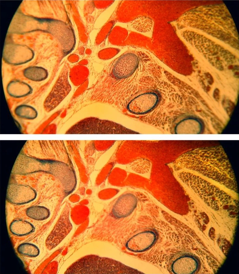
In entsprechender Weise konnte der selektive Autofokus bei gleichbleibender Fokussierung des Präparates wahlweise zentrale oder periphere Bereiche des Sehfeldes fokussieren, wenn für Testzwecke ein nicht plan korrigiertes Objektiv (einfacher Achromat) mit erheblicher Randunschärfe eingesetzt wurde (Abb. 12). Auch wenn man bereits manuell die Schärfenebene in etwa fokussiert hatte und die Fokussierung unverändert ließ, konnte man im Bildschirmbild mittels des selektiven Autofokus jederzeit eine Nachfokussierung interessierender Details vornehmen, von der Bildmitte bis zur Peripherie.