Die Mikroskopie-Kameras von The Imaging Source liefern Bilder für die digitale Pathologie
Veröffentlicht am 14. Januar 2021 von TIS Marketing.
Bei einer Reihe von schweren Blutkrankheiten wie Leukämie, Multiplem Myelom und Lymphomen ist die Differenzialzählung der Blutzellen in Knochenmarkausstrichen der diagnostische Königsweg. Gegenwärtig werden diese morphologischen Bewertungen noch manuell von Pathologen und anderen hochqualifizierten Labormitarbeitern durchgeführt und erfordern von den ausführenden Technikern ein hohes Maß an Konzentration und Präzision. "Human Factors" wie Stress, Ermüdung, Ablenkung und Ausbildungsstand können die Interpretation solcher Tests fehleranfällig machen, oder wie es die Wissenschaftler selbst nennen, "inter-operator variation". Um die Genauigkeit und Effizienz in der diagnostischen Hämatologie zu verbessern, hat aetherAI Microscope x Hema entwickelt - ein komplettes digitales Pathologiesystem, das die USB 3.0-Farbmikroskopiekameras von The Imaging Source nutzt, um digitale Bilder zu erstellen, die dann mit Deep Learning-Techniken verarbeitet werden.
Zellklassifizierung durch Deep Learning
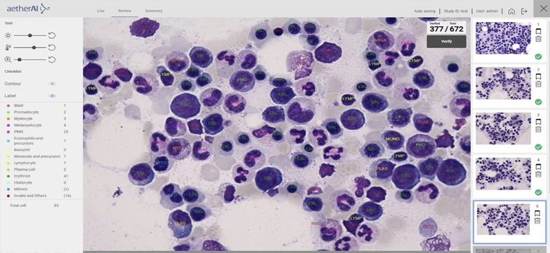
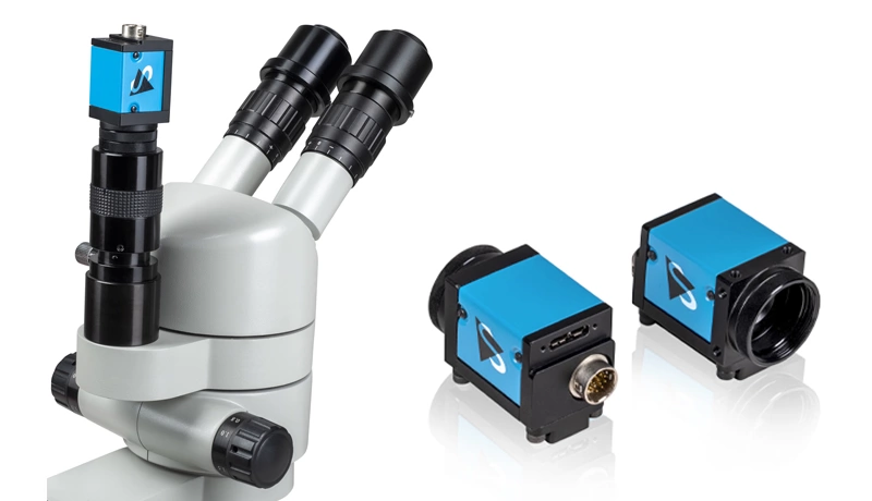
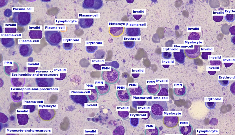
Um die CNNs des Systems richtig zu trainieren, arbeitete das Unternehmen mit dem National Taiwan University Hospital zusammen, um das weltweit erste KI-Modell für die Differenzialzählung von Knochenmarkabstrichen zu entwickeln. Das Modell wurde anhand eines umfassenden Bilddatensatzes von 500.000 kommentierten Knochenmarksproben trainiert. Die eingebettete Lösung von Microscope x Hema umfasst eine KI-gestützte Mikroskopsteuerungssoftware, ein KI-Modell für die Differenzialzählung und spezielle Hardware zur Unterstützung der KI-Inferenzierung. Bilder, die mit optischen Standardmikroskopen aufgenommen werden, enthalten oft komplexe Hintergründe, die sich negativ auf eine effiziente Zellanalyse auswirken können. Der hochempfindliche 20-MP-CMOS-Sensor der DFK 33UX183 Mikroskopkameras liefert rauscharme Bilder (hohes Signal-Rausch-Verhältnis). Die Bildvorverarbeitung der Kameras reduziert jegliches Restrauschen, um die Kanten und Konturen des Bildes zu verbessern, die Details hervorzuheben und die Bildunschärfe zu reduzieren. Die Bildalgorithmen von Microscope x Hema extrahieren Merkmale aus den Bildern und stellen dann Parameter wie Form, Kontur, unregelmäßige Fragmente, Farbe und Textur ein. Der Arbeitsablauf ist abgeschlossen, sobald das System die Zellen in der Probe klassifiziert und gezählt hat.
Durch die Entlastung des medizinischen Personals will aetherAI die Qualität der medizinischen Diagnostik verbessern, indem "Lösungen für die digitale Pathologie und KI-gestützte Diagnoseunterstützung angeboten werden." Unternehmensgründer Dr. Joe Yeh erklärte: "Die KI-Revolution wird die hohe Bedeutung digitaler medizinischer Bilder realisieren und das Gesundheitswesen auf die nächste Stufe bringen."