Microscope Camera: Comparison with Leica Camera
Published on August 22, 2016 by TIS Marketing.
Originally published in Mikroskopie in January 2016, this article was written by J. Piper and M. Torzewski. The English translation, written by Amy Groth, was serialized into: 1, 2, 3, 4, 5, 6, 7, 8, 9, 10 and 11.
Comparison with a Leica Add-on Camera
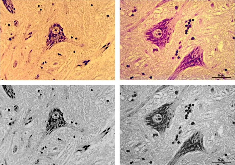
Even though a direct comparison of two different cameras can be problematic because of the diverse parameters and factors which may influence the results, nevertheless a comparison between the test subject of this article and the Leica MC 170 HD for orientation purposes should not be excluded. Using the Leica microscope DMLB, we started by photographing the neurons (in bright-field) of the specimens from Figs. 8, 9 and 11 with the aforementioned Leica system camera (using the microscope's original 40x Plan-corrected objective). The camera was affixed as designed by the manufacturer on the C-Mount head of the trinocular photo tube. The image displayed on the monitor had to be manually adjusted because the Leica camera is not equipped with One-Push autofocus. Subsequently for purposes of comparison, the same Plan-corrected objective was used with the camera / eyepiece combination from The Imaging Source to image the neurons of the same specimen (selective One Push autofocus was used here).
The Leica camera delivered a resolution of 2592 x 1807 pixels; the highest-possible resolution (4128 x 3096 pixels) was selected for the camera from The Imaging Source. The neurons were imaged significantly smaller by the later camera and its 10x eyepiece than in the case of the Leica camera and its special projective lens. Consequently, a section of the larger overall image (4128 x 3096 pixels) could be cut out which exactly corresponds to the original image size of the of the Leica camera (2592 x 1807 pixels); now both single images displayed the neurons in almost the same magnification. The panel of images in Fig. 17 juxtaposes the resultant images; in addition to the original color images, optimized black and white conversion images were also created. This comparison shows that for routine applications, such as the documentation of bright-field images, the camera from The Imaging Source need not fear comparison with a leading manufacturer's modular camera solution as it pertains to image quality.